Complications: Exposure
Exposure
The two most comprehensive studies to date reported very low rates of complications related to exposure in patients where the Bio-eye hydroxyapatite (HA) orbital implant was used. Shields’s (Shields 1993a) study of 200 cases revealed only 3 (1.5%) exposures, which were easily managed, and no cases of infection. Hornblass’s (Hornblass 1992) comprehensive survey of ophthalmic plastic surgeons, while it did not specifically cite a rate of exposure, did report rates of associated extrusions and infections of less than 2%. Traditional synthetic orbital implants, by contrast, were reported to have infection rates of 7 to 10%, and extrusion rates of 9 to 42% (Hornblass 1992). This excellent performance has made the Bio-eye HA implant the implant of choice for rehabilitation of the anophthalmic patient. The most common complication of the Bio-eye HA orbital implant is early exposure, i.e., an exposure occurring within the first few months postoperatively. This is believed to be related to surgical technique and not related to the chemical properties of the implant. The very coarse surface, because of its natural porosity, makes it critical that the surface of the implant be covered to prevent abrasion of the thin, overlying tissues. It takes approximately 6 months for a Bio-eye HA orbital implant to vascularize sufficiently to be fitted with a motility/support peg, at which time the implant is drilled to create a hole to accommodate the peg. Until the implant is vascularized, it should be treated as if it was any other orbital implant, i.e., any opening of the overlying conjunctiva and Tenon’s capsule should be avoided. Further, only a well-vascularized implant can accept a motility/support peg without undue risk of infection.
Preventing Exposure
The incidence of exposure can be greatly reduced by meticulous attention to surgical technique. Relatively few modifications of standard enucleation or evisceration techniques are needed to implant the Bio-eye HA orbital implant; but those modifications, while few, are crucial to achieving the best result.
Selecting Implant Size
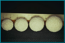
Table 1: Implant sizes required to fill orbits of various sizes.
An implant that is too large will cause tension on the overlying tissues and will increase the risk of exposure. Excessively large implants may also compromise the fornices and possibly limit motility. Also, an implant that is too large will not provide enough space for the ocularist to fashion an artificial eye with adequate (realistic) anterior chamber depth. Most ocularists can make a realistic artificial eye if they are provided with 3 to 5 mm of anterior-posterior thickness. It is also important to use the largest implant that the orbit can accommodate without causing the above-mentioned problems, since the volume of the globe should ideally be fully replaced by the prosthetic complex, which is comprised of the implant, artificial eye, and any implant-wrapping material used to cover the implant. Table 1 shows the implant sizes required to fill orbits of various sizes assuming that the average artificial eye has a volume of 2.5 cc).] It is important to objectively measure the size of the orbit using sizing spheres to ensure proper size selection, and to account for the volume added by use of a wrapping material. A scleral wrapping adds about 1.5 to 2.0 mm of thickness to the diameter of the implant. For example, if a 20-mm sizing sphere fits the orbit well, then an 18-mm sclera-wrapped implant should be used. In cases where an intermediate-size implant is needed, the anterior pole of a larger implant can be shaved down (flattened) with a scalpel to achieve the desired diameter and contour.
Wrapping the Implant
The surface of natural hydroxyapatite, from which the Bio-eye HA implant is made, is composed of numerous small spicules that may tend to abrade the overlying tissues as the implant moves. Since the conjunctiva and Tenon’s capsule are relatively thin tissues, this abrasion can easily lead to exposure. The risk of this type of exposure can be greatly reduced, in the case of enucleation, by wrapping the implant in sclera, fascia lata, or some other material prior to implantation. Naugle has suggested the use of tissue from the auricular muscle complex obtained from behind the ear (Naugle 1991) because of its proximity to the operative site, the familiarity of this area to ophthalmologists and ophthalmic plastic surgeons, and the inconspicuousness of the scar. In the case of fascia lata, Naugle has suggested obtaining the tissue from between the greater trochanter and the iliac crest, in order to avoid a conspicuous scar, herniation of the muscle belly, or hematoma formation (Naugle 1988).
Regardless of the material used, the implant should be wrapped in all cases except those in which compromised vascularity is suspected, such as in cases of prior irradiation treatment or where significant scarring has occurred. In these cases, a wrapping may unduly inhibit vascular ingrowth (Perry 1991) which is essential to the success of the implant. However, a small cap of wrapping material should be affixed over the anterior aspect of any bare implant in order to prevent abrasion of the overlying tissues. Alternatively, the rectus muscles can be sutured together over the anterior aspect of the implant–medial to lateral and inferior to superior. This layer of muscle tissue will provide an adequate barrier to protect the overlying Tenon’s and conjunctiva. Wrapping the Bio-eye HA implant also greatly facilitates placement of the implant deep into the orbit, since its rough surface does not catch on the soft tissue of the orbit during insertion. If a wrapping is used, a 10-mm hole should be cut in the material covering the posterior aspect of the implant to encourage more rapid vascularization. Do not place this uncovered area of HA anteriorly because of the increased risk of exposure. Windows should be cut in the sclera at the site of muscle attachment to put the muscle in contact with the implant and thus produce more rapid vascularization. Additionally, in the case of an enucleation, the sclera may be placed over the implant so that the corneal defect faces posteriorly (Dutton 1991).
Implant Placement
The Bio-eye HA orbital implant should be placed deep within the muscle cone and posterior to Tenon’s capsule (Soll 1972). An implant that is placed too far anteriorly may not provide adequate volume and will place undue tension on the overlying tissues. The fornices may also be compromised if the implant is placed too far anteriorly.
Proper Wound Closure
A common cause of early exposure is wound dehiscence at the suture line of Tenon’s capsule and the conjunctiva. These two tissue layers should be closed separately to provide the best barrier to exposure. This is especially important in cases where no wrapping material has been used to cover the implant (such as in cases of compromised vascularity of the orbital tissues following irradiation). Tenon’s capsule and the conjunctiva should be closed in separate layers with an interrupted 5-0 Vicryl suture. There should be absolutely no tension on the closure.
Selecting Conformer Size
Use the largest conformer that can be used without placing undue pressure on the fornices. A conformer that is too large will exert tension on the closure of the wound by exerting excessive pressure on the fornices. This is especially common in cases where the socket is contracted. The eyelids should close easily and completely with the conformer in place.
Allowing Vascularization
The Bio-eye HA orbital implant cannot fully deliver its unique benefits until it has vascularized. A well-vascularized implant resists exposure, extrusion, migration, and infection. Additionally, vascularization makes possible the use of a motility/support peg, which delivers all available movement directly to the artificial eye, while also supporting the weight of the eye, thereby preventing lower lid sag. Prior to placing the peg, the implant should be evaluated for vascularization via a bone scan or MRI with contrast. A poorly vascularized implant cannot support the epithelial lining that is needed to cover the interior of the peg hole.
Proper Fitting of the Artificial Eye
For optimum results, the artificial eye should be custom fitted using an impression technique. All edema of the socket must be allowed to subside completely before an accurate impression can be made. This usually takes 6-8 weeks post operatively. A poorly made prosthesis will place undue pressure on the tissues overlying the implant and may lead to pressure necrosis and eventual exposure.
Managing Exposures
Exposures are managed differently depending on their size, the time of their onset postoperatively, and the level of vascularity of the implant. Small exposures and those that occur later postoperatively may not require surgical intervention, while those that are larger and earlier, and those accompanied by significant discharge, should be managed surgically.
Premature Intervention
Recent reports have documented incidences of exposure (Beuttner 1992) and attempts to manage breakdowns of the conjunctiva as if they were exposures (Kim 1994). It should be noted that, especially with the Bio-eye HA orbital implant, conjunctival dehiscence does not constitute a frank exposure and, in most cases, does not require surgical intervention. Additionally, since the Bio-eye HA orbital implant can actually support the healing process from within the implant material itself, most small exposures tend to heal spontaneously. Most exposures occur within the first few weeks or months postoperatively, before the implant has had time to fully vascularize. It is not necessary to surgically intervene in order to manage all exposures with this implant, since most small exposures will heal spontaneously as the implant vascularizes. An implant that is well vascularized will heal more quickly than one that is poorly vascularized.
Early Exposures
If the exposure occurs within the first 6 months postoperatively, and if it is small (<= 3 mm), simply observe the exposure. If it widens, a graft should be considered. If the exposure does not widen and remains stable, continue to observe it, unless the patient complains of increased mucous discharge.
Late Exposures
If the exposure occurs later than 6 months postoperatively, and vascularization of the implant is confirmed, the exposure should simply be observed, unless the exposure widens or there is increased discharge. Most large exposures (>3 mm) will be accompanied by increased discharge and a graft is likely to be necessary. In some cases, even large exposures can remain stable for long periods of time if the implant is well vascularized.
Wound Freshening
In some cases, such as those in which the implant is adequately vascularized and the exposure is relatively small, it may be possible to encourage closure of the exposure by simply freshening the wound. The fibrous scar tissue of the conjunctiva/Tenon’s at the margins of the exposure should be burred to the point where viable vascular tissue can be seen. Blood should be noted at the margins of the exposure to ensure that viable tissue has been exposed by the burring. If the exposure does not close within several weeks after freshening the margins, a graft should be considered.
Implant Size Reduction
The HA material is easily shaped and can be reduced with the implant in the orbit, if necessary. In cases where the exposure is due to an over-sized implant, the anterior pole of the implant can be reduced by burring down the HA material through the exposure. Care should be taken to irrigate the area completely to remove any HA particles produced by the burring process. Copious irrigation with saline, followed by suctioning, is needed to remove any bacteria that may be in the pores due to the exposure. The implant should then be irrigated with antibiotics. After reduction of the implant is completed, the wound is closed without tension and a graft is used if necessary. The patient may also be placed on oral antibiotics postoperatively.
Grafting
In cases where surgical intervention is required, a graft should be considered. In no case should the exposure be closed by simply pulling conjunctival/Tenon’s tissue over the exposure with no barrier between the exposed hydroxyapatite spicules and the conjunctival/Tenon’s layer. Autologous dermis or hard palate serve well as graft materials for this purpose. Naugle has recently suggested the use of tissue from the auricular muscle complex as a patch graft (Naugle 1991). Vascularization of a graft may be enhanced by employing a conjunctival flap to provide an adequate blood supply to the graft material.
Vaulting the Artificial Eye
In cases where the artificial eye was fitted too soon or the fit was not accurate, there may be an exposure due to pressure necrosis. The artificial eye should only be fitted after edema in the socket has subsided, usually about 6 to 8 weeks postoperatively; otherwise, the posterior aspect of the eye will not match the shape of the socket and tissues overlying the implant. If pressure necrosis is suspected, the artificial eye should be vaulted over the area of the exposure or necrosis.
Conclusion
While the Bio-eye HA orbital implant represents a significant improvement over previous orbital implant designs, special care is required to achieve the full benefits inherent in the unique, natural hydroxyapatite material from which the implant is made. Relatively minor modifications of surgical techniques for evisceration and enucleation can greatly reduce the few complications associated with this implant.